Localization & Plasticity
 Localization of function is the theory that specific parts of the brain are responsible for specific behaviours or cognitive processes. The case study of HM is a good example of how a specific part of the brain has a specific function – that is, the hippocampus is responsible for transferring short-term memory to long-term memory. Although we know that some parts of the brain do play specific roles in behaviour, rarely does a part of the brain work in complete isolation. For example, in memory research, we argue that the cognitive process is the result of distributive processing rather than localization of function – in other words, several parts of the brain have to work together in order to help us create and retrieve memories. Today researchers continue to attempt to map the brain. One of the ways that they are doing this is by looking at the neural connections in the brain and creating a map called a connectome.
ATL: Inquiry
Star Trek episodes used to begin with the line, “Space – the final frontier!” But perhaps there is a frontier much closer to home – the human brain. One of the great challenges of the 21st century is to create a map of the brain that identifies the neural pathways of the brain and how they interact.
Go to the website for the “Human Connectome Project.” What do the researchers hope that this project will achieve? Why might this project be as “exciting” as the website claims?
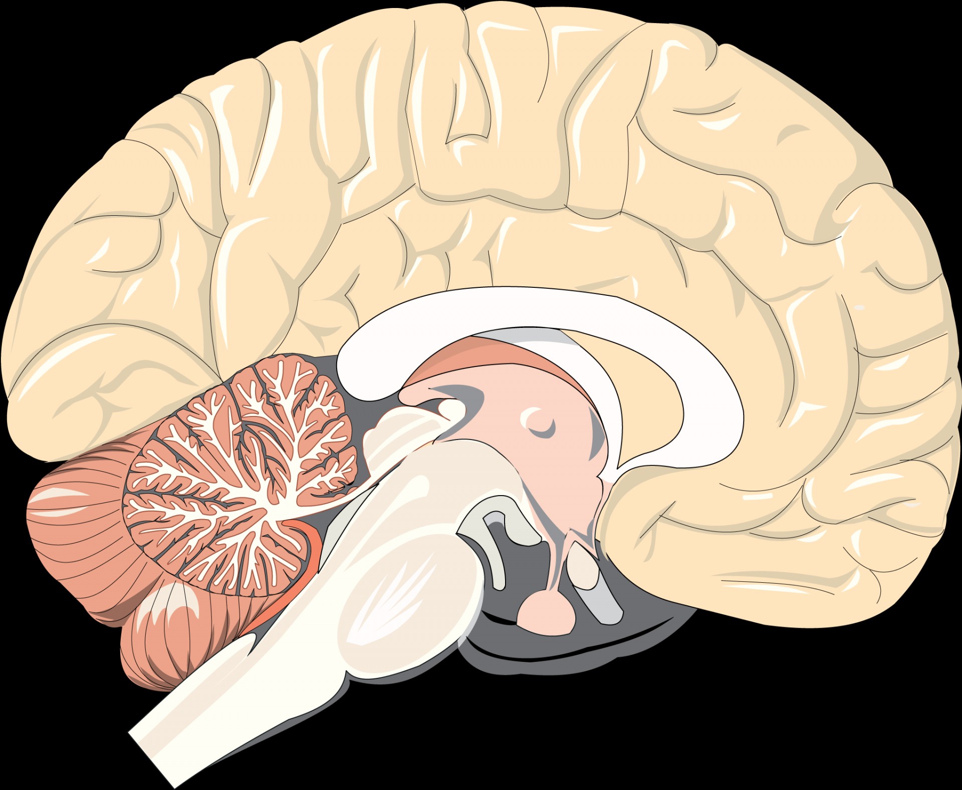
 When talking about the brain, we discuss four key areas: the brain stem, the cerebellum, the cerebrum, and the limbic system.
The brain stem is not usually studied by psychologists. The brain stem is responsible for regulating life functions, such as breathing, heart rate, and blood pressure. The cerebellum plays a key role in balance and motor function, including speech production. It also plays a role in learning – specifically, in classically conditioned responses.
The cerebral cortex is the largest part of the human brain, associated with higher brain function such as thought and action. The cerebrum is divided into four sections, called lobes.
Each of these lobes plays a key role in behaviour:
· The Frontal Lobe is associated with executive functions – that is, planning, decision-making, and speech.
· The Occipital Lobe is associated with visual processing.
· The Parietal Lobe is associated with the perception of stimuli;
· The Temporal Lobe is associated with auditory processing and memory.
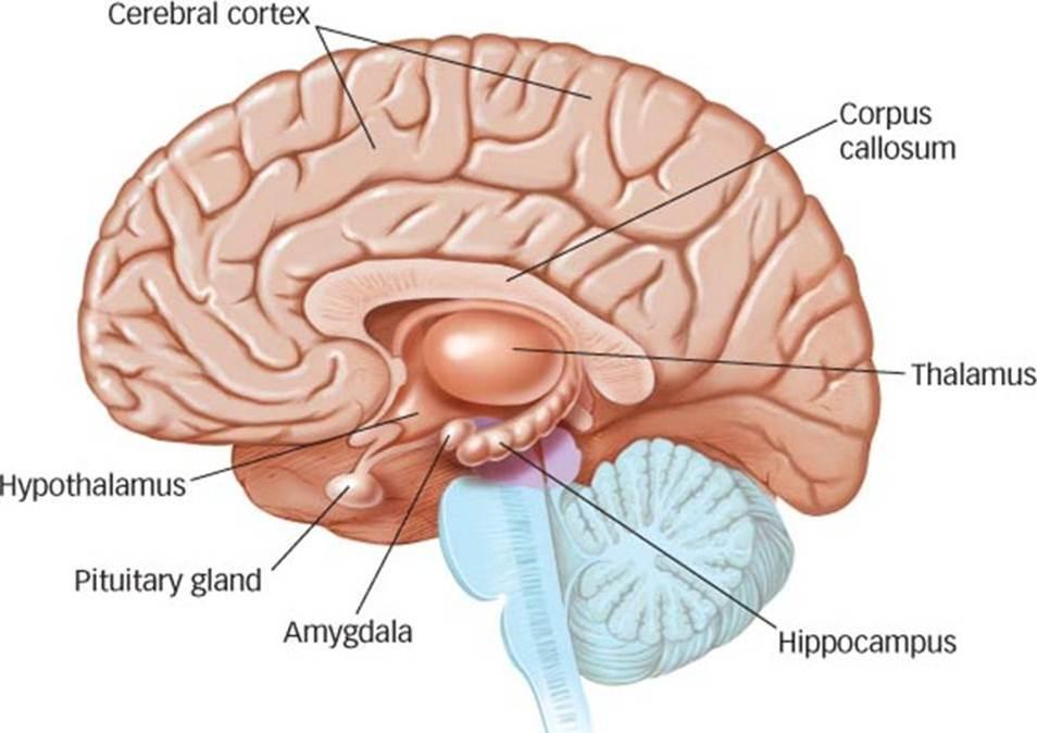
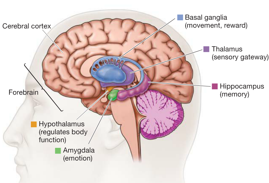
The Limbic System - often referred to as the emotional brain - is a major focus of psychological research for its role in memory and emotion. In the table below, you will find the key components of the limbic system.
The Limbic System
| Amygdala | Plays a role in the formation of emotional memory and fear responses. |
| Basal ganglia | Plays a role in habit-forming and procedural memory. |
| Hippocampus | Responsible for the transfer of short-term memory to long-term memory |
| Hypothalamus | Involved in homeostasis, emotion, thirst, hunger, circadian rhythms, and control of the autonomic nervous system. In addition, it controls the pituitary gland. |
| Nucleus accumbens | Plays a role in addiction and motivation. |
ATL: Communication
Choose one of the areas of the brain mentioned in this section and do a bit of research. Create a simple graphic that shows how the lobe or structure plays a role in human behaviour.
In the previous section, we looked at the case study of HM. Researchers were able to determine through their long-term study of HM that the hippocampus plays a key role in the transfer of information from short-term memory to long-term memory. A review of the study can be seen in the following video.
But as you can see, the answer to the question of how memory works was not complete from the study of HM. Other studies have shown us more about how memory works, including the case study of Eugene Pauly.
Research in psychology: The case of Eugene Pauly
 In 1992 Larry Squire and his team were introduced to one of the most interesting cases of amnesia since the famous HM study. At the age of 70, Eugene Pauly was diagnosed with viral encephalitis. Both his amygdala and hippocampus were completely destroyed. He demonstrated many of the same symptoms as HM.
Squire carried out a series of interviews with EP at his home. During one interview he asked EP to draw a map of his home. He was not able to do it. However, EP excused himself and got up to go to the toilet. How could a man that could not draw a map of his home find the bathroom on his own?
Several tasks that rely on procedural memories make this transition from involving an active frontal lobe to an active basal ganglia. Although the actual process is complex and not fully understood, when we are learning to do a task it is often cognitive in nature. So, when I first learn to drive a car, I need to think an awful lot about what I am doing. But remember, thinking takes up a lot of energy. Our brains have adapted in a way that minimizes the amount of energy that it needs to expend. Over time, the task is no longer cognitive, but what is referred to as an associative task. By chunking together a series of movements or behaviours, the task becomes automatic. The more common word we use for associative tasks is "habits."
EP was also able to take a walk around the block by himself. His wife would even follow him around the block to make sure that he was ok, but he was able to find his way home without any problems. When he was asked from any point on his walk where he lived, he would say he didn't know, but since the task was associative - or a habit - he was able to simply walk home. However, occasionally there was a problem. If the sidewalk was being repaired and he had to leave his familiar path, EP would get lost. Returning to our example of driving a car - even when we drive a long time, bad weather or heavy traffic forces us to concentrate more. The task reverts from associative to cognitive. When this happened to EP, he did not have the capacity to solve the problem as his memory was only procedural. Once the familiar pattern was changed, he was unable to complete the task.
In this case study, Squire & his team carried out several different research methods including interviews with EP and his family, psychometric testing (IQ testing), and observational studies. In addition, MRIs were used to determine the extent of the damage to EP's brain. MRI indicated that EP's basal ganglia were undamaged. It is believed that the basal ganglia are responsible for this type of procedural memory.
Click here for a complete description of the Case of Eugene Pauly.
Brain plasticity
The brain is a dynamic system that interacts with the environment. In a sense, the brain is physically sculpted by experience. Not only can the brain determine and change behaviour, but behaviour and environment can change the brain. Modern researchers argue that the brain is constantly changing as a result of experience throughout the lifespan.
Plasticity refers to the brain’s ability to alter its own structure following changes within the body or in the external environment.
Brain plasticity refers to the brain’s ability to rearrange the connections between its neurons - that is, the changes that occur in the structure of the brain as a result of learning or experience. High levels of stimulation and numerous learning opportunities lead to an increase in the density of neural connections. This means that the brain of an expert musician should have a thicker area in the cortex related to mastery of music when compared to the brain of a non-musician. The same can be said about students who spend a lot of time studying, compared to students who do not. Every time we learn something new, the neurons connect to create a new trace in the brain. This is called dendritic branching because the dendrites of the neurons grow in numbers and connect with other neurons.
This can be illustrated by a series of studies of brain plasticity carried out by Rosenzweig, Bennett, and Diamond (1972). The researchers conducted experiments where they placed rats into one of two environments to measure the effect of either enrichment or deprivation on the development of neurons in the cerebral cortex. In the enriched environment, rats were placed in cages with up to 11 other rats. In addition, there were stimulus objects for the rats to play with, as well as maze training. In the deprived environment, the rat was alone with no stimulation. The rats spent 30 or 60 days in their respective environments and then they were killed in order to measure the effect of the environment on their brain structures.
Post-mortem studies of their brains showed that those that had been in the stimulating environment had increased thickness in the cortex as a result of increased dendritic branching compared to the rats in the deprived environment. The frontal lobe, which in humans is associated with thinking, planning, and decision making, was heavier in the rats that had been in the stimulating environment. The combination of having "friends" to play with and many interesting toys created the best conditions for developing cerebral thickness.
This raises the question of the importance of stimulation and education in the growth of new synapses. If learning always results in an increase of dendritic branching, then the findings from animal studies that show increased dendritic branching in response to environmental stimulation are important for the human cortex as well. This was seen in a key study done by Maguire et al (2000). Unfortunately, environmental stressors can also have a negative effect on brain structures. Carrion et al (2009) found that children who had been abused tend to show a smaller hippocampus than their same-age peers.
Research in psychology: Maguire et al (2000)
 The aim of the study was to see whether the brains of London taxi drivers would be somehow different as a result of their exceptional knowledge of the city and the many hours that they spend behind the wheel navigating the streets of London. All potential taxi drivers must learn “the Knowledge” – that is, they must form a mental map of the city of London.
The participants for this quasi-experiment were 16 right-handed male London taxi drivers. The brain of the taxi drivers were MRI scanned and compared with the MRI scans of 50 right-handed males who did not drive taxis (the control group). In order to take part in the study, the participants had to have completed the "Knowledge" test and have their license for at least 1.5 years. The controls were taken from an MRI database. The sample included a range of ages so that age would not be a confounding variable.
The study is correlational as the IV was not manipulated by the researcher but naturally occurring. The researchers were looking to see if there was a relationship between the number of years of driving a taxi and the anatomy of one's brain. It was also a single-blind study - that is, the researcher did not know whether she was looking at the scan of a taxi driver or a control.
There were two key findings of the study. First, the posterior hippocampi of taxi drivers were significantly larger relative to those of control subjects and the anterior hippocampi were significantly smaller. Secondly, the volume of the right posterior hippocampi correlated with the amount of time spent as a taxi driver. No differences were observed in other parts of the brain. Maguire argues that this demonstrates that the hippocampus may change in response to environmental demands.
The study of localization of function and brain plasticity are very much linked. Studies from both abnormal and developmental psychology have demonstrated, for example, the way that stress has an effect on memory by interfering with the work of the hippocampus. In addition, long-term stress appears to lead to hippocampal atrophy - that is, hippocampal cell death that leads to a smaller hippocampus. This was found in a study by Bremner (2003).
Thirty-three women participated in this study, including women with early childhood sexual abuse and PTSD (N=10), women with abuse without PTSD (N=12), and women without abuse or PTSD (N=11). The researchers used an MRI to measure the volume of the hippocampus in all of the participants - and a PET scan to measure its level of function during a verbal declarative memory test. Women that were abused and showed symptoms of PTSD were found to have a 16% smaller volume of the hippocampus compared to women with abuse without PTSD. In addition, these women showed a lack of activity in the hippocampus when carrying out the memory task. Women with abuse and PTSD had a 19% smaller hippocampal volume relative to women without abuse or PTSD.
Synaptic plasticity
From a biological point of view, how does neuroplasticity happen? How is it possible that new synapses are formed? And how it is possible that synapses also decay?
According to biologists, synaptic plasticity works by the maxim: use it or you lose it!
Synapses become stronger through repeated use. This is known as long-term potentiation. LTP leads to a greater level of response – that is, longer periods of depolarization - on the post-synaptic membrane. Over time, this leads to protein synthesis and gene expression which will be the building blocks used for dendritic branching – a process called neural arborization.
When a synapse is not used or is under-stimulated, it may go through the process of synaptic pruning. It is believed that this is the way for the brain to remove synapses that are no longer needed, making the functioning of the neural networks more efficient. The process of pruning is still not fully understood.
If you are interested in an in-depth explanation of the process, you may want to watch the video below. Simply being able to link the above information to a study like Draganski (2004) is all you need to be able to do to answer an SAQ on Paper 1.
Exam preparation: Linking to curriculum
When discussing the localization of brain function and neuroplasticity, there are several other examples throughout the course that you may choose to use. Here are some examples.
- Localization of function: the case of HM and the hippocampus
- Localization of function: the case of Eugene Pauly and the basal ganglia
- Localization of function: the role of the amygdala in flashbulb memory.
- Localization of function: the role of the ventromedial prefrontal cortex in decision making
Checking for understanding
Which of the following is an example of localization of function?
When more than one part of the brain is involved, this is a distributive function. The change in the brain's structure in reaction to the environment is known as neuroplasticity. The different hemispheres of the brain do help the opposite side of the body; this is called in lateralization, not localization.
Which lobe is responsible for processing visual information?
Which of the following might be a symptom of damage to the frontal lobe?
If there was damage to the amygdala, which symptom might we observe?
Which part of the neuron is most affected by learning?
Neuoplasticity is the result of "dendritic branching" - and this is what happens when we learn through interacting with our environment.
Which of the following is not true of Rosenzweig, Bennett & Diamond's (1972) study?
The study does not do a good job of isolating variables. It is unclear if it was the visual stimulation, the social interaction with other rats or the exercise provided by a track wheel that led to the denser frontal lobe in the rats.
According to Carrion (2009), what effect can stress in the environment have on the brain of children?
Stress hormones lead to hippocampal cell death. This has an effect on the ability of children to form long-term memories, and thus is a sign of cognitive impairment.
What research method did Maguire use for her study of brain plasticity in taxi drivers?
There was no manipulation of the IV. The length of time that they were driving a taxi was not manipulated by the researcher and they could not be randomly allocated to groups. Since the could not be any demand characteristics from the participants as they could not change the structure of their brains, the study does not need any deception. However, in order to prevent researcher bias in interpreting the structure of the brain, the researchers were unaware of which brain they were examining. This means it is a single-blind control.

 IB Docs (2) Team
