 IB Docs (2) Team

Methodology: biological approach
Over the past thirty years, few areas of psychology have developed quite so rapidly as brain research. As technology has progressed, so has our ability to monitor and map out the brain’s activity. The brain is seen as the control center of human functioning. Psychologists seek to understand how the brain matures over a lifetime and how it adapts to the environment.
Prior to the development of modern scanning technology, one of the most common ways to study the brain was through the use of case studies of brain damage. Often such studies provide researchers with a situation that they could never ethically reproduce in a laboratory. Case studies of brain-damaged patients are often carried out longitudinally - that is, over a long period of time - in order to observe both the short-term and long-term effects of damage.
In addition, case studies are often holistic in their approach, looking at a range of effects of the damage, rather than a single behaviour. In order to do this, case studies use triangulation. In the case of a patient with brain damage, this may include interviews with the family, psychometric testing – for example, IQ or personality testing, experiments and observations.
However, case studies have limitations. First, as the researchers do not manipulate an independent variable, no cause and effect relationship can be determined. In addition, since case studies are of single individuals, from a single case study we cannot generalize the findings to all human beings. And since it is naturally occurring, the study cannot be replicated. Finally, it may be very difficult to verify information about the patient before the accident took place. Information about the individual’s IQ, problem-solving skills, memory or interpersonal skills is often reliant on the memories of family members. In addition to the fact that this is not an accurate measure, the memories of the family members may not be accurate.
Data triangulation: When more than one source of data is used. For example, a case study of schools that looked at stress in the IB program in five different schools. In this way, if we get consistent findings, we know that it was not simply because of the school that we chose.
Method triangulation: When more than one research method is used. If we get consistent findings, that means that the choice of research method was not the reason for our findings.
Researcher triangulation: When more than one researcher studies a case. Researchers are able to compare their observations and interpretations in order to increase reliability and credibility.
Theory triangulation: When we look at a case from more than one theoretical perspective - e.g. biological, cognitive, and/or sociocultural.
In spite of these methodological limitations of a single case study, psychologists agree that they provide important information that can be used to study the effects of brain damage over time, as well as spark new research. Psychologists do not use a single case study to draw definitive conclusions about the role of the brain on behaviour. Instead, they often use similar cases to verify their findings and they may even perform experiments with animals to investigate the hypothesized relationship between damage to a specific area of the brain and behaviour. If there are several case studies of damage to the same area of the brain that show the same consequences on behaviour, biologists may conclude that damage to a specific part of the brain may cause a specific behaviour. This is the case with the famous case study of HM which has contributed to our understanding of the relationship between the brain and memory.
Research in psychology: The case study of HM
 HM fell off his bicycle when he was aged 7 and sustained a serious head injury. Epileptic attacks began when he was 10. At the age of 27 he had become so incapacitated by his seizures that he could not lead a normal life and medication did not help him. With the approval of the patient and his family, neurosurgeon William Scoville performed an experimental surgery where he removed tissue from the medial temporal lobe (including the hippocampus) on both sides of HM’s brain.
After the operation, HM remembered his childhood very well. His personality appeared largely unchanged. However, HM suffered from anterograde amnesia – he was no longer able to transfer information from short-term memory to long-term memory.
In order to carry out her research, Milner used many different strategies. This is an example of how method triangulation may be used in a case study:
- Psychometric testing: IQ testing was given to HM. His results were slightly above average with an IQ of 104 before the operation and a slightly improved IQ of 112 after the operation due to the reduction in seizures.
- Direct observation of his behaviour;
- Interviews with both HM and with family members.
- Cognitive testing: memory recall tests as well as learning tasks - such as reverse mirror drawing.
- Corkin (1997) later did an MRI to determine the extent of the damage done to HM's brain.
HM could not acquire new episodic knowledge (memory for events) and he could not acquire new semantic knowledge (general knowledge about the world). This suggests that the brain structures that were removed from his brain are important for long-term explicit memory.
Memories in the form of motor skills, i.e. procedural memories, were well maintained, for example, he knew how to mow a lawn. He also showed improvements in the performance of new skills such as reverse mirror-drawing in which he had to acquire new eye-hand coordination.
An MRI scan of HM’s brain was performed in 1997 and 2002 where Corkin analyzed the extent of the damage. It was possible to see that parts of HM’s temporal lobe including the hippocampus had the most damage. Damage to the hippocampus explains the problem of transferring short-term memory to long-term memory.
The researchers concluded that the hippocampus plays a critical role in converting memories of experiences from short-term memory to long-term memory. Since HM was able to retain some memories for events that happened long before his surgery it indicates that the hippocampus is not the site of permanent storage but rather plays a role in the organization and permanent storage of memories elsewhere in the brain.
(Scoville & Milner, 1957; Milner & Klein, 2015)
TOK: Ethics
When discussing the HM case study, there are several important questions about the ethics of the study. For example, even though his parents gave consent for the research, is this acceptable, considering that HM was not able to give consent himself? Although HM’s identity was kept confidential, was the longitudinal study of his life intrusive?
Is it acceptable to argue that since we learned very important information about the brain from this research that the research is justified in spite of our ethical concerns?
The use of technology in brain research
 Many early experiments on the brain involved invasive techniques—for example, removing (ablation) or scarring (lesioning) brain tissue in animals in order to study subsequent behavioural changes. Behaviour before and after lesioning was compared. In a classic study, Hetherington and Ranson (1942) lesioned a part of the brain called the ventromedial hypothalamus (VHM) in rats, a part of the brain that is believed to control feeding. As a result of the lesioning, the rats increased their food intake dramatically and often doubled their weight. This led researchers to conclude that the hypothalamus acted as a brake on eating.
The use of invasive techniques raises serious ethical concerns and psychologists have to conform to codes of conduct for the use of non-human animals; for example, the Ethical Principles of Psychologists and Code of Conduct by the American Association of Psychologists. This means that psychologists who engage in research with animals need to apply to an ethical committee for permission to carry out their research. If the potential harm to the animal cannot be determined, permission will only be granted if the research is considered to add to current knowledge and there are no alternatives.
Modern technology is now extensively used in psychology because it provides an opportunity to study not only brain structures but also the active brain while avoiding many of the ethical concerns of animal experimentation. This allows researchers to see localization of function – that is, the functions of specific parts of the brain and how they relate to behaviour.
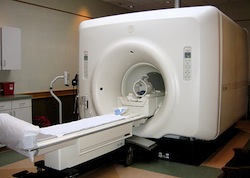
One imaging technique is Magnetic Resonance Imaging [MRI]. The MRI gives a three-dimensional picture of the brain structures. The MRI was used in the case study of HM to determine the extent of his brain damage. An MRI scanner uses a magnetic field and radio waves to map the activity of hydrogen molecules, which are present in different brain tissue to different degrees. The image can either be viewed as a slice of the brain from any angle, or it can be used to create a three-dimensional image of the brain.
There are several advantages of using an MRI scan. First, the procedure is non-invasive, with minimal potential harm to the participant. Secondly, the image has high resolution; this gives researchers a good sense of the actual structure of the brain. However, the MRI only indicates structure; it does not actually map what is happening in the brain. In addition, MRI research is correlational in nature, not allowing researchers to establish a clear cause and effect relationship.
In order to observe the activity of the brain, other technologies are used. Positron Emission Tomography – commonly known as PET scanning - is used to observe metabolic processes in the brain by detecting the gamma rays emitted indirectly by a tracer. PET neuroimaging is based on the assumption that areas of high radioactivity are associated with brain activity. Before a PET scan begins, a patient is given a safe dose of a radioactive tracer compound introduced into the body by a modified glucose molecule [FDG]. The injected FDG enters the bloodstream, where it can travel to the brain. If a particular area of the brain is more active, more glucose will be needed there. When more glucose is used, the radioactive tracer is detected by the PET scanner. The scan, which usually takes between 30 minutes and two hours, produces a multi-coloured image that shows which parts of the brain were the most active. The colour of each dot shows the intensity of the energy signal.
One of the key advantages of the PET scan is that it allows participants to perform psychological tasks while the researcher observes brain activity. There are two key limitations of PET scanning. First, it requires an injection with a small amount of radioactive material.. Although this will not cause harm to the participant, it is still an invasive practice and raises ethical concerns. Secondly, PET scanning is quite slow and has relatively poor resolution. So, although it does indicate where brain activity is taking place, it is not as clear as more modern technology like the fMRI.
Unlike the MRI, which shows the structure of the brain, an fMRI (functional magnetic resonance imaging) shows actual brain activity and indicates which areas of the brain are active when engaged in a behaviour or cognitive process. The fMRI measures changes in blood flow in the active brain. These scans have a higher resolution than PET scans, and they are easier to carry out. This is one of the most frequently used technologies in biopsychological research today. It tracks changes in blood flow and oxygen level as a measurement of neural activity. When a specific brain area is active, it uses more oxygen, and therefore the blood flow increases. This can be detected by the fMRI scanner.
Unlike PET scanning, fMRIs are non-invasive. There is no radioactive isotope necessary. The quality of the image is also much better and rather than a static image, the fMRI produces a film that demonstrates a change in the brain over the period of the scan.
For both the MRI and the fMRI, there are certain precautions that must be taken to protect the safety of the participant. Since the technology works with a powerful magnet, it is important that objects that contain iron be removed. In rare cases, a participant may have to be removed from a sample because of a metal implant – for example, certain types of pacemakers or cochlear implants.
The fMRI scanner is not a natural environment for cognition. Therefore, research may lack ecological validity. There is a lot of noise in the tunnel and participants may experience anxiety due to the claustrophobic nature of the machine. There is a question of artifacts in the imaging – that is, some of the activity may be related to anxiety or reaction to the machine, rather than the behaviour that is being studied.
The use of colors may exaggerate the activity of the brain. Much of the activity of the brain is spontaneous and is not a reaction to stimuli. Therefore, it is difficult to know exactly which areas of the brain are active in a behaviour.
Brain areas activate for various reasons – just because the amygdala lights up, doesn’t mean that fear is necessarily part of the response being observed.
ATL: Thinking critically
The use of PET and fMRI scans has helped psychologists to identify brain patterns for dysfunctional behaviours. In fact, some scientists say that these scanning images are like fingerprints. There is a certain pattern for people with schizophrenia, alcoholism, and depression, among other disorders. These patterns are present, even if the person does not show any symptoms of the disorder.
You see in the news an article that says that your government is considering having all members of government who need security clearance have a PET or fMRI scan in order to determine whether they are “fit” for this position. As a psychologist, how would you respond to this suggestion?
You see in the news an article that says that your government is considering having all members of government who need security clearance have a PET or fMRI scan in order to determine whether they are “fit” for this position. As a psychologist, how would you respond to this suggestion?
Students should be able to discuss the pros and cons of using brain imaging technology in predicting future behaviour. Although there may be some trends in negative behaviors with regard to brain damage, the predictive validity of brain imaging technology is limited and highly controversial.
Very often, students will be completely against the use of brain scanning. You may want to have them read the following articles as a way of extending the discussion.
Throughout the course, you will find several studies that use brain imaging technology. Because of that, they are not described in this section. In revising for exams, you may want to consider the following studies.
MRI | fMRI |
Checking for understanding
Which of the following is not a characteristic of a case study?
Case studies cannot be replicated. They often presente unique situations; however, more importantly, because they are longitudinal (taking place over time) and are holistic (looking at many aspects of an individual) and do not follow a standardized procedure, they are not possible to replicate. Similar case studies can be used to see if the results present a trend. Credibility -- or the validity of the study - is established by seeing if the same results are obtained using different methods.
Which of the following is an example of triangulation?
Triangulation means to use two or more research methods, sources of data or research methods. Having more than one researcher is known as researcher triangulation. By comparing the data that they collect, we can see whether the variable being measured is reliable. This is often known as inter-rater reliability or inter-coder reliability.
To what extent is it possible to generalize from a single case study?
Which of the following is not a conclusion that we can draw from the HM study?
It appears that memory is not stored in the hippocampus, but the hippocampus transfer information from short-term to long-term memory and then moves information from long-term memory to short-term memory.
Which brain imaging technique could be considered invasive?
In order to carry out a PET scan, participants are injected with a radioactive isotope. This would be considered an invasive technique.
Which of the following is true about the use of brain imaging techniques in the study of brain?
Anxiety or fear can lead to artefacts - which may indicate brain activity that is irrelevant to what is being studied. The colors do not tell us what is actually happening, only where there is some activity taking place in the brain. The scanner is a very unnatural environment, so for an fMRI scan, there is the question of ecological validity. However, since the MRI only looks at structure, ecological validity is not relevant. A scan can be easily replicated; even though it is rather expensive to do so.
What is a key difference between and MRI and an fMRI?
The modern MRI and fMRI are the same machine, just different functions. The MRI is a static image of the brain whereas the fMRI is like a film of the functioning brain.
 Twitter
 Facebook
 LinkedIn