Yoghurt and milk bacteria experiment
 This lab is a safe way to demonstrate the structure of prokaryotes and to introduce the variety of roles which prokaryotes have in metabolism. The comparison of slides of milk, yoghurt and kefir illustrates the variety of bacteria and their small size compared to eukaryote cells, like fungi. The usefulness of staining in microscopy can also been seen, using Gram staining or Methylene blue. This lab could be used as a 'mini IA' to introduce students to one or more aspects of the Individual assessment, such as sample size or hypothesis testing.
Note: The latest IB guidance on use of microorganisms in the IA advises against growing microbes from classroom surfaces and with covid-19 protocols raising awareness of hygiene this lab is a safe introduction to prokaryote shape and size.
Lesson Description
 Guiding Questions
Is it prokaryotes or eukaryotes which turns milk into curd, yoghurt or kefir?
What challenges will need to be overcome to see any prokaryotes?
Activity 1 - Chemicals in milk and yoghurt making
Watch the Sci show video and answer the questions on the student worksheet which helps to make links between molecules like proteins in milk and the difference between milk and yoghurt from a chemical sense. There is also an introduction to cell structure and protein structures.
Answer the questions on the ![]() Questions about milk and yoghurt sheet below.
Questions about milk and yoghurt sheet below.
Activity 2 - Making yoghurt in the the kitchen or the lab
Carry out the experiment ![]() Making yoghurt in the kitchen or the lab.
Making yoghurt in the kitchen or the lab.
This could be used as preparation for Activity 3 Gram staining bacteria in yoghurt, the yoghurt will take about 24 hours to set at 30°C.
For further information and see the Wikihow to make yoghurt
Activity 3 - Staining and viewing bacteria in yoghurt.
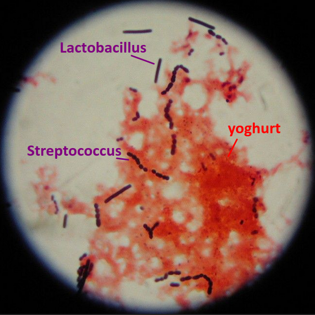
This activity includes three methods to view bacteria in a microscope capable of x400 magnification or higher. The first is a simple fresh slide with no staining, the second involves fixing the sampel to the slide and adding Methyline blue stain, and the third illustrates staining with the more complex Gram staining method. These methods are not required for IB diploma Biology but they may be useful for the Extended essay, or for student IAs.
Carry out one method or more from the ![]() Experiments to stain and identify yoghurt bacteria worksheet, shown below.
Experiments to stain and identify yoghurt bacteria worksheet, shown below.
Teacher's notes
In recent years there have been frequent comments in the DP Biology Examiner's reports about use of microorganisms.
The guidance now states clearly that in IA's students should only culture known non-pathogenic strains of bacteria, and advises that students should not swab door handles or hands.
This protocol using yoghurt bacteria can be said to use 'known non-pathogenic strains', yoghurt bought for human consumption has been tested during production. This greatly reduces the risk of culturing pathogenic bacteria, however this does not eliminate it completely, so the usual strict rules of hygiene and aseptic technique should be used.
This video from microbehunter gives a nice commentary on the experience of making a wet mount slide of yoghurt using a slide and coverslip, without staining. After some searching he finds streptococcus bacteria at x400 magnification, and he also has a lot of protein / yoghurt clumps as students will find.
If the experiment doesn't work well in the lab, this video would be a little view into the viewing of bacteria.
- Why? - the big picture. Prokaryotes are small but have a different size and structure of cells to humans, or other eukaryotes.
- Hook - Why do we eat so many bacteria? - the video in Activity 1. There are links to other parts of the IB DP Biology course such as protein structure and denaturing of proteins in the worksheet from Activity 1
- Equip / Experience the main facts / skills
- Milk as food contains casein and lactase, mostly, lactose structure, respiration, lactase enzymes, etc.
- gram staining colours, prokaryote shapes and names like bacillus and coccus, steptococcus can be used in microbiology.
- Rethink - shift perspective to enhance understanding. Use the data collected and the information in Activity 3 to identify different species of bacteria and try to identify them.
- Tailor - personalise the learning / differentiation. There are options in the use of the three activities, and it may be preferable for some students to begin with Activity 3 and Gram's staining, other's may prefer the theory of milk protein, and the links to theory first. To save time Activity 2 can be omitted and yoghurt can be bought from a shop.
- Organise the sequence. It's up to you to choose how to sequence this activity. Some might like to begin with making yoghurt, then look at the theory in Activity 1 and in the following lesson to complete Activity 3. Others may prefer to put everything into one lesson.
Further information
Yoghurt lab protocolhttps://mymission.lamission.edu/userdata/brownst/docs/Yogurt%20Preparation%20Lab.pdf
A great simple method to view yoghurt bacteria from Microscope.com. microbe lab
https://www.microscope.com/bacterial-succession-in-milk/
Simple yoghurt bacteria and description of shape: https://www2.mrc-lmb.cam.ac.uk/microscopes4schools/yoghurt.php

 IB Docs (2) Team
