Alveoli & gas exchange
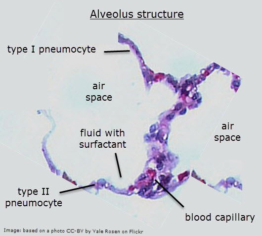
Students learn about the structure of the lungs and their alveoli including type I and II pneumocytes. After a short quiz on basic lung structure a short screen cast helps students to complete a diagram of the cells of two alveoli and a capillary. This diagram would be suitable for an answer during an IB exam. Using flashcards students' learn the diagram labels and annotations of their function. A final test of the concepts through some IB style questions with answers further consolidates learning.
Lesson Description
Guiding Questions
What is the difference between ventilation, gas exchange and respiration?
How many different cell types are found in the alveolus?
Which parts of the alveoli are responsible for gas exchange?
Activity 1 Drawing lung diagrams
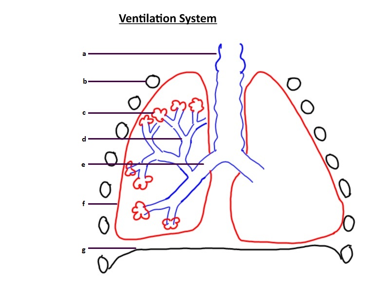
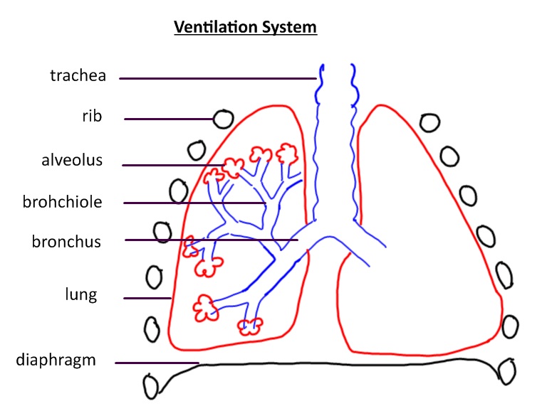
It is assumed that students already know the general structure of the lungs. Here is a little test.

Click the eye icon for the labels

Play this short screencast showing a way to draw the pneumocyes of alveoli.
 |
Complete the ![]() alveolus diagram activity sheet while you watch. Replay or pause the video as you wish.
alveolus diagram activity sheet while you watch. Replay or pause the video as you wish.
There is a ![]() detailed diagram of the lungs including alveoli here for reference, but of course it is too complex to draw in an IB exam.
detailed diagram of the lungs including alveoli here for reference, but of course it is too complex to draw in an IB exam.
Activity 2 Understanding the structure & learning the labels
Study the eleven Flash cards and then take the self-marking online test.
![]() Screencast - Ways of usng these Flashcards in a lesson (4 mins)
Screencast - Ways of usng these Flashcards in a lesson (4 mins)
Activity 3 - IB style questions
Complete the ![]() IB style questions below to really test understanding of these structures.
IB style questions below to really test understanding of these structures.
Teachers' notes
This page covers the new requirement of students to understand the structure of alveoli is more details. In particular the presence of type I and type II pneumocytes.
The simple diagram shown in the screen cast is designed specially to be easy enough for students to replicate under exam conditions.
A more detailed diagram link is always given to support understanding and raise awareness of the beauty and complexity of biological systems. There are some fabulous images of pneumocyte cells on the Yale university website.
Some students can learn a diagram and it's labels easily, but many students need help to remember the precise shapes and sizes of the parts of each diagrams, and they need practice to get the labels correct and to understand the labels. The resources on this page aim to help teachers support these students.
There are many ways to use this page.
- Students can work independently or as a group.
- The resources can be projected in the classroom or studied on a computer.
- Worksheets allow you to print as much or as little as you wish.
- Differentiation is possible, as students could follow their own learning pathways to the IB style questions.
A Suggested Lesson Plan.
Aims: To learn the features of alveoli and how to draw a diagram of the ventilation system
Starter: 10 minutes
- Students complete the Diagram Activity worksheet while watching the Screencast as a whole class.
Main: 40 minutes
- Students study printed (or online) Flashcards to help memorizing of the labels
- Students take a Quick test - to see how well they understand the labels
- Students review the Screencast and attempt the diagram (with labels) from memory.
End: 10 minutes
- Plenary - students look at slides of cells to further consolidate understanding of alveolus structure
- Differentiation - students who need additional time for main can continue independently.
Homework
Complete the IB style questions.
Answers are available on a separate page
- (choose whether or not to allow students to see this page in the Student access page
Extra resources
- An excellent description of gas exchange Respiratory Tract Histology by Dr A McLeod
- Another super explanation of the Histology of the lungs from Leeds University
- A university talk through of some structures in the alveoli
- A clear wiki commons diagram of cells in an alveolus by Katherinebutler1331 CC-BY-SA-4.0
- Nice histology slides from trachea to alveoli through the ventilation system by John McNulty at LUMEN.

 IB Docs (2) Team

