Brain structure and function
 The human brain rapidly develops from the neural tube and a hugely complex organ results. In this activity students answer some questions about basic processes involved in brain formation. Activity 2 reinforces an understanding of the structure and function of the brain using a great resource from NINDS and the final activity looks in a little detail at the sympathetic and parasympathetic branches of the autonomic nervous system.
Lesson Description
 Guiding Questions
How can a neural tube structure develop into a brain and spinal cord?
What are the main structures found in the brain called?
What is the general function of each structure?
Activity 1 - Brain formation
Early brain formation is the formation of the neural tube.
What follows is the expansion of the anterior part of the neural tube (the part nearest the head)
Quote from the Mayo Clinic:
"The neural tube is the embryonic structure that eventually develops into the baby's brain and spinal cord and the tissues that enclose them. Normally, the neural tube forms early in the pregnancy and closes by the 28th day after conception. In babies with spina bifida, a portion of the neural tube fails to close properly, causing defects in the spinal cord and in the bones of the backbone."
Look through these slides and then answer the questions below.
Questions
This slide shows a timeline of neural tube development.
The embryonic brain develops complexity through enlargements of the neural tube called vesicles.
- The primary vesicle stage has three regions,(28 days)
- The secondary vesicle stage has five regions. (35 days)

1) Describe the changes to each of the coloured parts of the neural tube after 35 days.
..................................................................................................................................................
..................................................................................................................................................
2) Describe what must be happening to individual neurones within these structures
..................................................................................................................................................
..................................................................................................................................................
3) What effect will these developments have on energy requirements of a growing embryo?
..................................................................................................................................................
Click the eye icon for model answers.
Model answers
1) Describe the changes to each of the coloured parts of the neural tube.
The spinal cord differentiates from the lower part of the neural tube. It gets longer, and it could show spina bifida if the tube hasn't properly formed.
The top, brown vesicle becomes the cerebral cortex
The second vesicle (orange) becomes the hypothalamus and other structures.
The third vesicle (green) becomes midbrain.
The fourth (pink vesicle) becomes the cerebellum [and the Pons - not in the IB syllabus
The fifth region becomes the medulla oblongata.
2) Describe what must be happening to individual neurones within these structures
As a fetus grows, nerve cells (neurons) are produced and they travel to their eventual locations within the brain.
The survival of any one neuron is not guaranteed and those neurones that do not find a place where they can live and thrive — are destroyed by neural pruning.
3) What effect will these developments have on energy requirements of a growing embryo?
Watch this ![]() Two minute brain development video from Neuroscientifically Challenged to illustrate how the brain develops from the anterior part of the neural tube
Two minute brain development video from Neuroscientifically Challenged to illustrate how the brain develops from the anterior part of the neural tube
Note that:
- The brain develops from the anterior part of the neural tube.
- The cerebral cortex forms a larger proportion of the brain.
- Extensive folding of the cerebral cortex helps it to fit into the cranium.
- Neurons are initially produced by in the neural tube.
- Immature neurons migrate to a final location as they mature
None of the 'cephalon' names are required for IB.
Click the eye icon to the see the video on the page.
Activity 2 - Brain structures
In activity 1 the names of the brain structures required in the IB guide have been introduced.
Read the following booklet ![]() MINDS - Know your brain to find the functions of the following parts of the brain:
MINDS - Know your brain to find the functions of the following parts of the brain:
- medulla oblongata, in the brain stem
- cerebellum,
- hypothalamus,
- pituitary gland, and
- cerebral hemispheres
- Visual cortex,
- Broca’s area,
- nucleus accumbens ( you may need to Google this one)
Complete ![]() Brain structure and function activity sheet to record the structures, their functions and how you think experiments, lesions and fMRI scans have helped study brain function Use of animal experiments, autopsy, lesions and fMRI to identify the role of different brain parts.
Brain structure and function activity sheet to record the structures, their functions and how you think experiments, lesions and fMRI scans have helped study brain function Use of animal experiments, autopsy, lesions and fMRI to identify the role of different brain parts.
.
Activity 3 - The autonomic nervous system
The brain controls automatic responses using the autonomic nervous system.sympathetic and parasympathetic antagonistic effects)
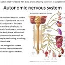
The autonomic nervous system consists of peripheral neurons which originate in the brain stem and the spinal cord. It controls ‘involuntary’ processes in the body, those which don’t require conscious control, for example; swallowing, breathing and heart rate.
It is an antagonistic system because it has two branches which have opposite effects.
Sympathetic (Fight or flight) | Parasympathetic (Rest & digest) |
Dilates pupils Decreases salivation Increases release of glucose Prevents activity in digestive system Increases heart rate | Constricts pupils Increases salivation Conserves body energy Increase gut activity Slows down heart rate |
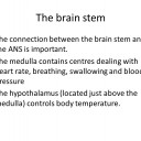
The connection between the brain stem and the ANS is important. The medulla contains centres dealing with heart rate, breathing, swallowing and blood pressure while the hypothalamus (located just above the medulla) controls body temperature.
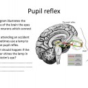
An example is the pupil reflex
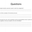
Complete the following worksheet of questions: ![]() Autonomic nervous system
Autonomic nervous system
Teacher's notes
Activity 1 is based on a small point on the IB guide, but it is interesting. Don't spend too long on this activity, students only need a general understanding of these details.
For those students who like to know more, this is a nice clear explanation of the neural tube development:![]() Development of the human brain from factheworld
Development of the human brain from factheworld
This is another nice brain link Take a 3-D tour of the brain
Key points from the IB guide:
- The brain is formed from expansion of the anterior part of the neural tube.
- Brain metabolism requires a lot of energy.
Activity 2 covers the parts of the brain and some of the methods biologists have used to uncover the functions of different parts of the brain.
Activity 3 covers the autonomic nervous system and how it controls involuntary processes in the body, for example swallowing, breathing and heart rate. The questions can be used as a worksheet or in class using the slides.

 IB Docs (2) Team

