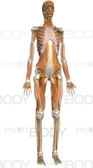
Muscles & joints
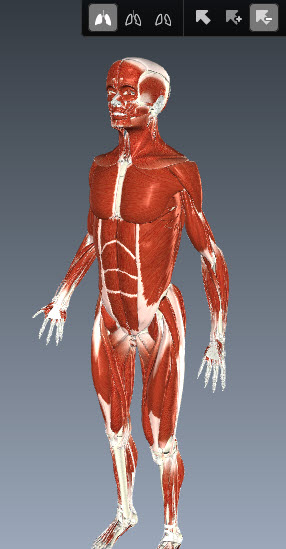
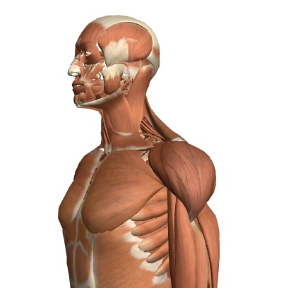 Using a model skeleton or interactive web tools like "Zygotebody" and "Visible Human" students identify different joints in the skeleton before exploring the detail of synovial joints and the human elbow. There are questions to answer and a worksheet to support student record keeping.
Lesson Description
Guiding Questions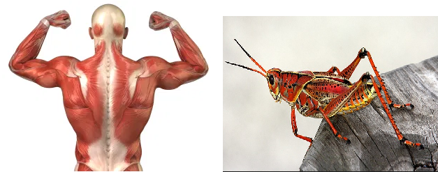
What is the difference between a bone and an exoskeleton?
Explain how they are similar in the way they interact with muscles to create movement.
Activity 1 - Finding different types of join on the skeleton
The best way to find the different joints in the human skeleton is by investigating a model skeleton. In the absence of a real model there are some great interactive human bodies online. Zygote body and Biodigital human are both free to use, but they both require a login.
Explore one of the interactive bodies to answer the questions below.
Questions
- Describe two similarities between the elbow and the knee joint.
- Distinguish two differences between the elbow and the knee.
- What structures do Ligaments attach to?
- Compare and contrast the hip joint with the elbow joint.
Activity 2: Labelling the parts of a synovial joint
There is a nice section of Biodigital Human which explains common disorders of the elbow joint.
This makes a nice introduction to the structure of the elbow. A radiology masterclass of the elbow.
(The old link seems not to be working at the moment: //bit.ly/1zxogro)
- Discuss how the elbow joint works and compare the hip and knee
- Complete Human Joints worksheet
Activity 3 - Exoskeletons, muscle contraction and insect legs
This activity looks at the structure of a grasshopper leg or a crabs claw (not an insect, but bigger and easier to see). Students learn about antagonistic muscle attachments and similarities in the way that muscles operate in insects.
Basic information about how grasshopper legs work Click the image / link to find out about muscles and elastic tendons in a grasshopper leg. | Video experiment to measure grasshopper jump force. |
Teachers notes
In activity 1 students are best directed to simply find different types of joints between bones in the skeleton. This can be effectively done using a model skeleton in the lab but two online interactive human bodies are suggested in this activity as an alternative. These have the advantage of giving a view of ligaments, tendons and muscles as well as bones.
The questions in this activity guide students in their research and they could be answered using screen shots of the online skeleton images.
Activity 2 is focused on the elbow joint. There is a worksheet to support note taking but a text book would be useful for answering the questions given.
A detailed set of radiology and mri scan images can be found here.
A nice youtube illustration of the elbow here
Activity 3 is an introduction to insect leg structure and muscles in insects.
Extension work - Introduction to the molecular details of muscle contraction
The next lesson is likely to be about the sliding filament theory. Watch the following animation to get an overview of sliding filament theory and a preview of the next lesson.

 IB Docs (2) Team

