 IB Docs (2) Team

The eye and photoreceptors
 This activity begins with an interesting case of academic honesty from the 1930s. The structure of the retina is discovered and students learn how to annotate retina diagrams. A short second activity give some practice at labelling parts of the eye and a final activity investigates optic chiasma to consolidate student understanding.
Lesson Description
 Guiding Questions
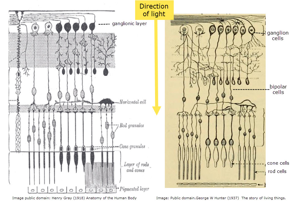
These two diagrams of the cells of the retina were published in separate books 20 years apart.
1) Do you think there is any case for a inquiry about 'academic honesty'?
2) Can you see any differences in the representations of:
- cone cells
- rod cells
- bipolar cells
- ganglion cells?
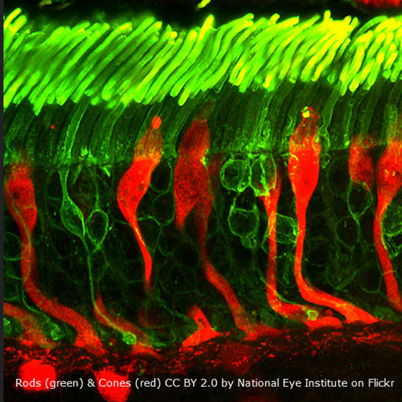
Activity 1 - Rod and cone cells of the retina
Identify the components of this retina image.
Annotations must show - direction of light, rod cells, cone cells, bipolar cells and ganglion cells.
The following image shows the cells of the retina, light would enter on the left.!
Drag the text boxes below to tag the labels A to E:
Bipolar cell Cone cell Rod cell Motor neuroneGanglion cell
|
A. B. C. D.
|
.jpg)
This is a slightly unusual diagram as the light enters the diagram from the left. Notice how a group of rod cells connect to a bipolar cell, but only one cone cell.
Key points about the retina
- Rods and cones are photoreceptors located in the retina.
- Rod cells are sensitive to low light levels and any wavelength.
- Cone cells require higher light intensities and they are only sensitive to specific wavelengths of light.
- Bipolar cells send the impulses from rods and cones to ganglion cells.
- Ganglion cells send messages to the brain via the optic nerve
Activity 2 - Structure of the eye
Complete ![]() The structure of the eye worksheet shown below.
The structure of the eye worksheet shown below.
The diagram of human eye should include the sclera, cornea, conjunctiva, eyelid, choroid, aqueous humour, pupil, lens, iris, vitreous humour, retina, fovea, optic nerve and blind spot.
Activity 3 - Fields of view in the visual cortex
The information from the right field of vision from both eyes is sent to the left part of the visual cortex and vice versa.
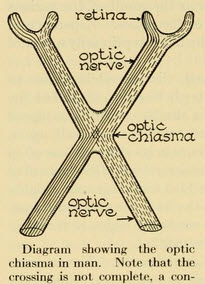
Look carefully at the diagram above by George W. Hunter and answer the following questions.
Questions
1. The visual cortex is found at the ends of the optic nerve, partly in the left hemisphere and partly in the right hemisphere of the brain. From which parts of the retinas of the eyes do the nerves leading to the left hemisphere visual cortex originate?
.......................................................................................................................................................................
2. Explain why the right hand side of your field of vision forms images on the left side of each retina.
.......................................................................................................................................................................
.......................................................................................................................................................................
.......................................................................................................................................................................
3. It is thought that this arrangement of neurones in the optic chasma helps to allow 3D (Sterioscopic) vision.
Why would it facilitate the comparison of images from both right and left eyes in order to estimate the distance of an object?
.......................................................................................................................................................................
.......................................................................................................................................................................
4. Interestingly vegetarian Xenopus tadpoles don't have overlapping fields of view, and all the neurones from each eye go to the opposite hemisphere of the brain. As the Xenopus tadpoles metamorphose into carnivorous African clawed frogs the neurones cross as in the arrangement shown above.
Suggest a reason for this change.
.......................................................................................................................................................................
.......................................................................................................................................................................
.......................................................................................................................................................................
Click the eye icon to display model answers
Model answers
1. The visual cortex is found at the ends of the optic nerve, partly in the left hemisphere and partly in the right hemisphere of the brain. From which parts of the retinas of the eyes do the nerves leading to the left hemisphere visual cortex originate?
The left side of each retina.
2. Explain why the right hand side of your field of vision forms images on the left side of each retina.
The lens in each eye focuses the light on the retina,
The pupil is a small hole in the front of each eye through which light passes.
So light from the right travels in a straight line through the pupil and the lens, and falls on the left side of each retina.
3. It is thought that this arrangement of neurones in the optic chasma helps to allow 3D (Sterioscopic) vision.
Why would it facilitate the comparison of images from both right and left eyes in order to estimate the distance of an object?
An object in the right field of view will form two images on the left sides of each retina. The eye must compare these images to estimate the distance of the object. This comparison will be more rapid if the neurones involved are all found in the same location of the cerebral cortex.
4. Interestingly vegetarian Xenopus tadpoles don't have overlapping fields of view, and all the neurones from each eye go to the opposite hemisphere of the brain. As the Xenopus tadpoles metamorphose into carnivorous African clawed frogs the neurones cross as in the arrangement shown above.
Suggest a reason for this change.
It is thought that carnivores who must catch their prey have an advantage if they have overlapping fields of view and 3D vision.
Tadpoles are vegetarian, have eyes on the sides of their head and don't have 3D vision.
It is logical that as the eyes turn forwards during metamorphosis, creating 3D vision, that the arrangement above should form in the optic chiasma.
How the maturing neurons can migrate and produce this change is surely one of the wonders of nature.

Teacher's notes
Activities to help students understand the structure of the retina and the eye form the most part of this activity 1 and 2.
Activity 3 tries to address the interesting arrangement of neurons in the optic nerve, leading to the visual cortex.
*****
These extra materials are not ready yet, but they may appear later.
- Edge enhancement & Contralateral processing
- Application: Red-green colour-blindness as a variant of normal trichromatic vision.
 Twitter
 Facebook
 LinkedIn