Option A - Neurobiology SL Revision list
This pages gives outline details of the content of Option A for SL students. There are essential questions and lists of student skills and applications. Helpful for revision. Student can check their understanding and prioritise areas for revision . 
A.1 Neural development
- Embryonic chordates form a neural tube by in-folding of the ectoderm followed by elongation of the tube formed.
- Differentiation of cells in the neural tube produces neurons.
- Immature neurons migrate to their final location.
- Each immature neuron grows an axon in response to chemical stimuli.
- Some axons extend out of the neural tube to reach other parts of the body.
- Note: Terminology relating to embryonic brain areas or nervous system divisions is not required.
- Multiple synapses are formed by developing neurons.
- Synapses that are not used do not persist.
- Neural pruning involves the loss of unused neurons.
- The nervous system has plasticity, the ability to change with experience.
Revision Question(s)
- How does the nervous system form in a developing embryo?
- Which cells are the first to become nerve cells?
- How do the cells organise themselves to form a nervous system.
- What is the role of connections between neurones in the development of the nervous system?
- What happens to unused synapses?
- What happens to synapses which are used regularly?
Skills ( can you ...)
- Understand how brain function may be reorganized after a stroke.
- Apply an understanding of closure of the embryonic neural tube to the condition spina bifida in which the tube closure is incomplete..
- Annotate a diagram of embryonic tissues in Xenopus, used as an animal model, during neurulation
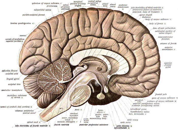
A2 The Human Brain
- The brain is formed from expansion of the anterior part of the neural tube.
- Brain metabolism requires a lot of energy.
- Skill: Identification of parts of the brain in a photograph, diagram or scan of the brain, including the medulla oblongata, cerebellum, hypothalamus, pituitary gland and cerebral hemispheres.
- The brain stem contains the autonomic nervous system which controls involuntary processes in the body. For example: Swallowing, breathing and heart rate coordinated by the medulla.
- Application: Use of the pupil reflex to evaluate brain damage.
The cerebral cortex is larger (in proportion) and more highly developed in humans than other animals.- An increase in the total area of the cerebral cortex has extensive folding so it fits within the cranium.
- The cerebral hemispheres are responsible for higher order functions.
- Different parts of the brain have specific functions. For example; Visual cortex, Broca’s area and nucleus accumbens.
- Application: Use of animal experiments, autopsy, lesions and fMRI to identify the role of different brain parts.
- The left cerebral hemisphere receives sensory input from sensory receptors in the right side of the body and the right side of the visual field in both eyes and vice versa for the right hemisphere.
- The left cerebral hemisphere controls muscle contraction in the right side of the body and vice versa for the right hemisphere.
Revision Question(s)
How can a neural tube structure develop into a brain and spinal cord?
What are the main structures found in the brain called?
What is the general function of each structure?
- How do biologists find out about different structures of the brain?
- Why does an large and extensively folded cortex in humans give humans better higher order thinking skills than animals without so many folds?
- What are higher order thinking skills?
- Is it true that the left hemisphere controls the right side of the body?
Skills ( can you ...)
- Appreciate that exceptions occur to any rule, for example the brain can even reorganize itself following a disturbance such as a stroke.(It is the rule that specific functions can be attributed to certain areas, brain imagery shows that some activities are spread in many areas)
- Look at electroencephalogram images and recognise abnormal patterns that can be used to diagnose conditions, e.g. Angelman syndrome, which is a genetically inherited condition.
- Analyse correlations between body size and brain size in different animals.
A3 Perception of stimuli
- Receptors detect changes in the environment. e.g. Humans sensory receptors - mechanoreceptors, chemoreceptors, thermoreceptors and photoreceptors.
- Application: Detection of chemicals in the air by the many different olfactory receptors.
- Rods and cones are photoreceptors located in the retina - they differ in their sensitivities to light intensities and wavelengths.
- Bipolar cells send the impulses from rods and cones to ganglion cells.
- Ganglion cells send messages to the brain via the optic nerve.
- The information from the right field of vision from both eyes is sent to the left part of the visual cortex and vice versa. (covered in the cerebral cortex work in section A2.)
- Structures in the middle ear transmit and amplify sound.
- Sensory hairs of the cochlea detect sounds of specific frequency.
- Impulses caused by sound perception are transmitted to the brain via the auditory nerve.
- Hair cells in the semicircular canals detect movement of the head.
Revision Question(s)
- 5% of your genes code for chemoreceptors in the nose. Why is the sense of smell so important in animals?
- Chemoreceptors have receptor molecules, but how do the photoreceptor cells of the retina detect changes in the environment?
- What are the four different types of receptor?
- What are the two types of photoreceptor in the eye and how do they differ?
- through which neurones are the photoreceptors connected to the brain?
- How do the eyes' left and right fields of view connect to the visual cortex?
Skills ( can you ...)
- Label a diagram of the structure of the human eye - including sclera, cornea, conjunctiva, eyelid, choroid, aqueous humour, pupil, lens, iris, vitreous humour, retina, fovea, optic nerve and blind spot.
- Annotate a diagram of the retina recognising cell types - rod and cone cells, bipolar neurons and ganglion cells and the direction in which light moves.
- Use your knowledge of the retina to explain red-green colour-blindness as a variant of normal trichromatic vision.
- Label the structure of the human ear, including pinna, eardrum, bones of the middle ear, oval window, round window, semicircular canals, auditory nerve and cochlea.
- Apply your knowledge of the cochlea to explain the use of cochlear implants in deaf patients.

 IB Docs (2) Team
