Drawing the Human Kidney
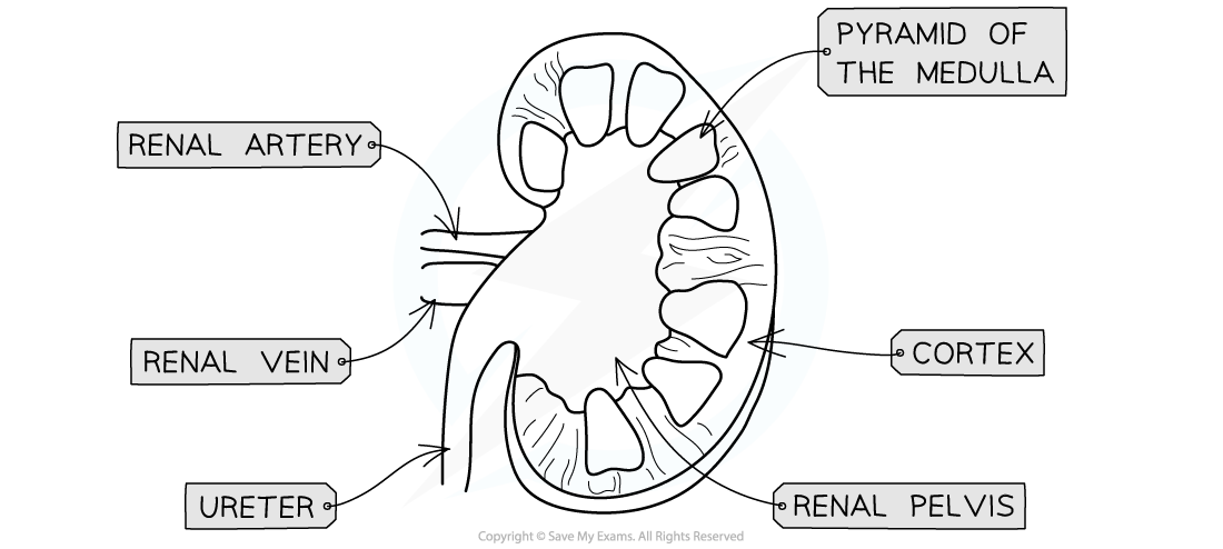
- The following elements should be included in a drawing of the human kidney
- The kidney should be roughly oval in shape with an inward curve on the side where it is connected to the blood vessels and ureter
- The renal vein should be wider in diameter than the renal artery
- All three kidney regions should be clearly shown
- An outer cortex that takes up roughly a fifth of the kidney’s diameter
- The medulla within the cortex
- The medulla has regions called ‘pyramids’ which should be visible
- The renal pelvis on the inward curve of the kidney’s side, and draining into the ureter
- Lines should be drawn in clear, dark pencil with no sketching
- Label lines should not cross each other and should cross as little of the drawing as possible
- No shading or colour is required

A drawing of the kidney should be an accurate representation of the kidney’s shape, show the three main regions of the kidney, and show the kidney’s connections to blood vessels and the ureter
Annotating the Nephron
- Each kidney contains thousands of tiny tubes, or tubules, known as nephrons
- Nephrons are the functional unit of the kidney and are responsible for the formation of urine
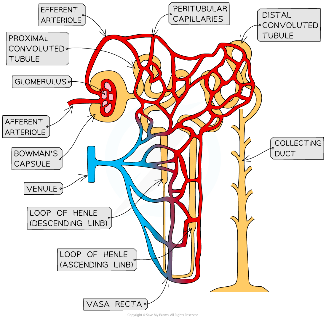
- An annotated diagram of the nephron should include the following elements
- The glomerulus
- A ball of capillaries inside which blood flows at high pressure to bring about ultrafiltration
- The afferent and efferent arterioles of the glomerulus
- The afferent arteriole brings blood into the glomerulus from the renal artery and the efferent arteriole carries blood away from the glomerulus and into the peritubular capillaries
- The afferent arteriole is wider than the efferent arteriole to generate high blood pressure in the glomerulus
- Remember, a comes before e in the alphabet, and afferent comes before efferent in the blood flow through the kidney
- The Bowman’s capsule
- A cup-like structure into which the blood of the glomerulus is filtered
- The proximal convoluted tubule
- The section of the kidney tubule in which most reabsorption of sugars, water, and salts occurs
- It is lined with tightly packed cells that have microvilli and many mitochondria to aid the reabsorption process
- Remember, proximity means that something is close by, so the proximal convoluted tubule is closest to the glomerulus
- The loop of Henle
- A hairpin-like structure that concentrates the filtrate and creates a concentration gradient across the medulla in order to maximise water reabsorption
- The descending limb descends into the medulla, while the ascending limb ascends back up into the cortex
- The distal convoluted tubule
- The section of tubule that connects the loop of Henle with the collecting duct; more water is reabsorbed here
- The amount of water reabsorbed depends on the amount of ADH in the blood
- Remember, distant refers to something that is far away, so the distal convoluted tubule is furthest away from the glomerulus
- The collecting duct
- This tube descends back into the medulla and drains into the renal pelvis
- More water is reabsorbed here
- The amount of water reabsorbed depends on the amount of ADH in the blood
- The vasa recta
- The capillary that flows alongside the loop of Henle and into which water and salts are reabsorbed
- Peritubular capillaries and venules
- Peritubular capillaries are a network of blood vessels in close proximity to the nephron into which substances are reabsorbed
- Venules carry blood back towards the renal vein

Nephrons are the functional unit of the kidney
