Analysing Muscle Contractions in Electron Micrographs
- Many biological structures are too small to be seen by the naked eye
- Optical microscopes are an invaluable tool for scientists as they allow for tissues, cells and organelles to be seen and studied
- Electron microscopes provide a much higher magnification and resolution so sub-cellular details can be studied
- Using microscopes to calculate the size of specimens requires the use of an eyepiece graticule which should be calibrated to the microscope
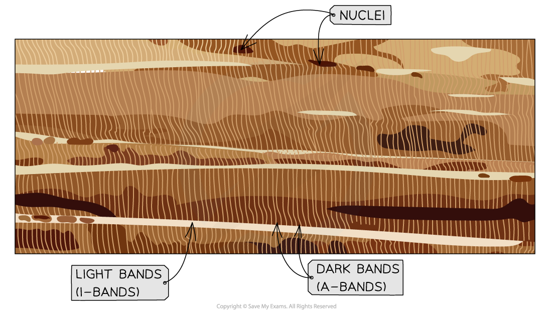
- However, it can be very difficult to make out the features of skeletal muscle fibres using an optical microscope
- Only the banding is visible, this is why it is referred to as striated muscle

The dark bands produce a characteristic striped appearance under an optical microscope
- Electron microscopes are often used to see muscle fibres in more detail
- They reveal the structure of myofibrils

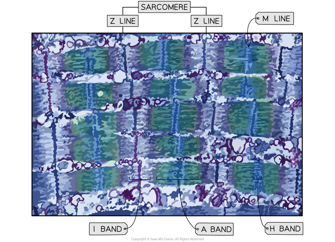
 The detailed structures of the muscle fibres are visible due to the much stronger magnification of the electron microscope.
In a relaxed sarcomere:
- There will be visible dark lines where the Z-lines are at either end of the sarcomere
- There will also be a darker band in the middle of the sarcomere where the thicker myosin fibres are positioned and in the very centre of that is the M line
- Around the Z-line, lighter bands are seen where the thinner actin fibres are positioned
In a contracted sarcomere:
- The Z-lines and M-lines are still visible with a shorter distance between the two z-lines
- The lighter bands around the z-line will be smaller or not visible
- The darker band will be the same size (although may appear a bit darker).
