Skeletal Muscle Fibres: Structure
- Muscles in the body that are attached to the skeleton and aid movement are called skeletal muscles
- Other muscle types include:
- Cardiac muscle which is found in the heart
- Smooth muscle is found in the blood vessels and organs
- Skeletal muscle is striated as it has a stripy appearance when viewed under a microscope
- Striated muscle cells are bundled up into fibres which are surrounded by a single plasma membrane called the sarcolemma
- The fibres are highly specialised cell-like units
- Each muscle fibre contains:
- An organised arrangement of contractile proteins in the cytoplasm
- Many nuclei – this is why muscle fibres are not usually referred to as cells
- Specialised endoplasmic reticulum called the sarcoplasmic reticulum (SR) which stores calcium and conveys signals to all parts of the fibre at once using protein pumps in the membranes
- Specialised cytoplasm called the sarcoplasm contains mitochondria and myofibrils
- The mitochondria carry out aerobic respiration to generate the ATP required for muscle contraction
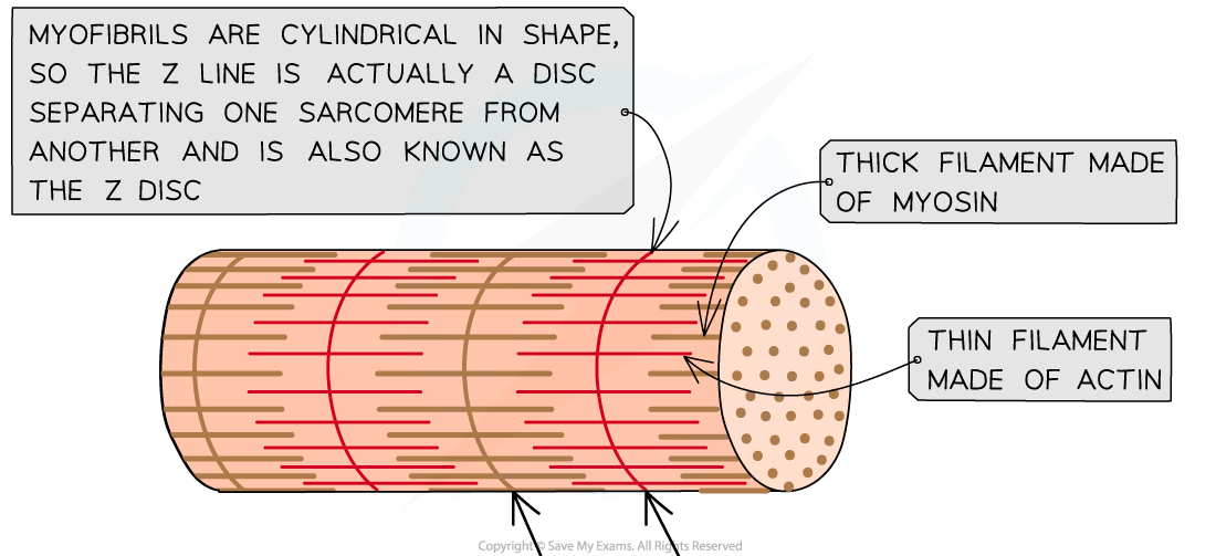
- Myofibrils are bundles of actin and myosin filaments, which slide past each other during muscle contraction
- Each muscle fibre contains:
- The sarcolemma (muscle fibre membrane) has many deep tube-like projections that fold in from its outer surface
- These are known as transverse system tubules or T-tubules
- These run close to the SR


The ultrastructure of striated muscle and of a section of muscle fibre
Myofibrils
- Myofibrils are located in the sarcoplasm
- Each myofibril is made up of two types of protein filament:
- Thick filaments made of myosin
- Thin filaments made of actin
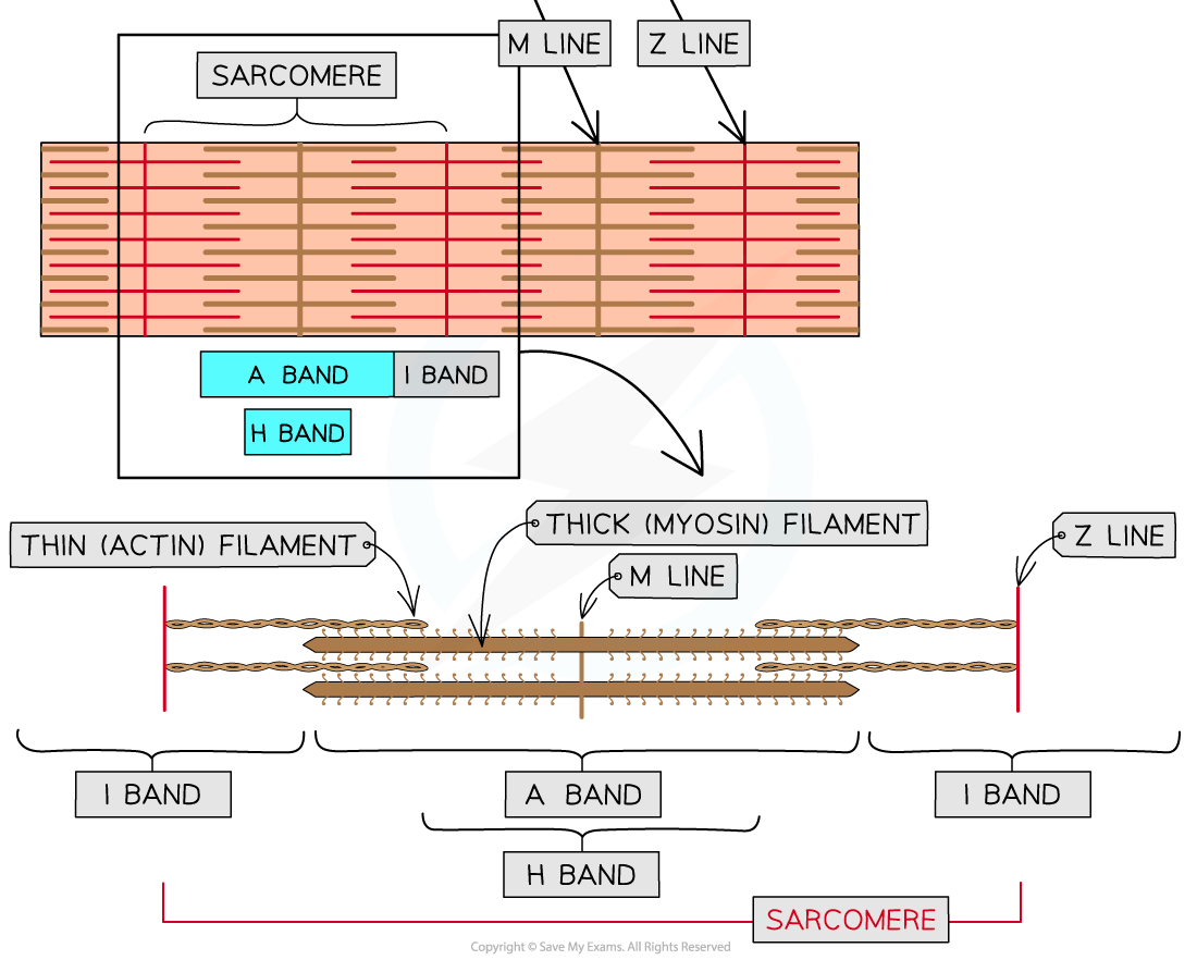
- These two types of filament are arranged in a particular order, creating different types of bands and lines
Myofibrils Parts & Descriptions Table


The structure of a myofibril
