History of Fluid Mosaic Model
NOS: Using models as representations of the real world; there are alternative models of membrane structure
- Scientists use models to represent real world ideas, organisms, processes and systems that cannot be easily investigated. Scientists can experiment on the models enabling them to test predictions and develop explanations for observations made
- Over time as technological developments have been made the models used to represent the structure of membranes of cells and organelles have changed
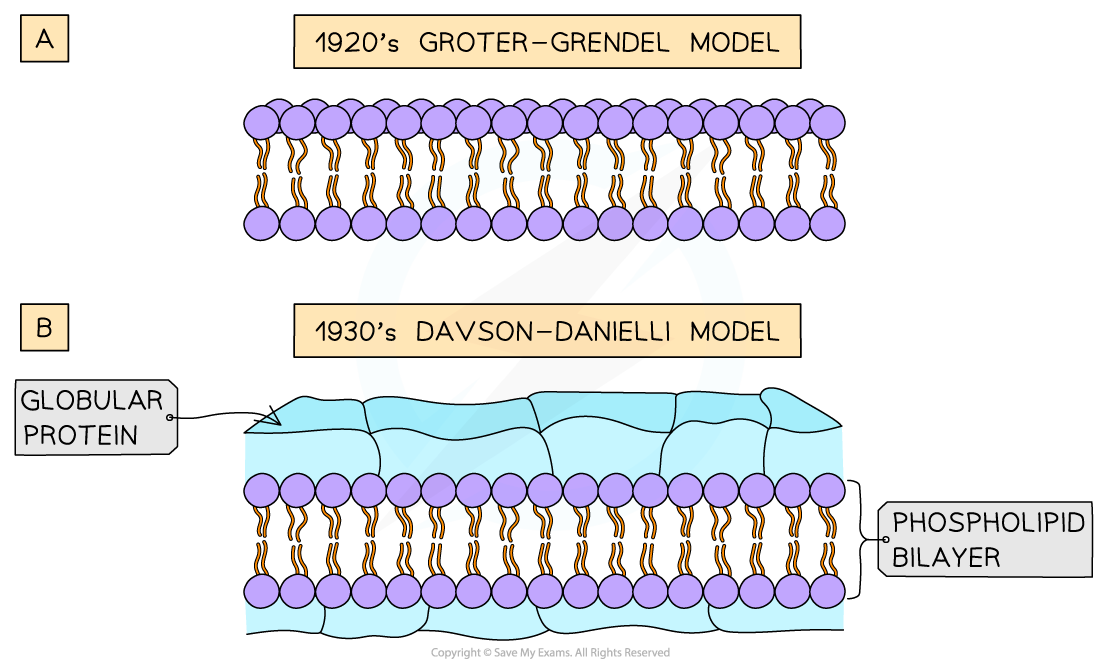
1920's Gorter and Grendel
- The Gorter and Grendel model showed that the phospholipids in the membrane of cells were arranged into a bilayer
- Evidence for this model:
- The number of phospholipids extracted from red blood cell membranes was double the area of the plasma membrane if it was arranged as a monolayer
- Problems with this model:
- Their model did not explain the location of proteins or how molecules that were insoluble in lipids moved into and out of the cell
1930's Davson and Danielli
- Davson and Danielli's model of the membrane suggested that the proteins were arranged in layers above and below the phospholipid bilayer
- Evidence for this model:
- Membranes were effective at controlling the movement of substances in and out of cells
- Electron micrographs showed the membrane had two dark lines with a lighter band between. In electron micrographs, proteins appear darker than phospholipids
- Problems with this model:
- Freeze-etched electron micrographs of the centre of the membrane showed globular structures scattered throughout
- Improvements in technology used to analyse the proteins in the membranes showed that proteins were globular, varied in size and had parts that were hydrophobic
- These problems suggested it was unlikely that the proteins would form continuous layers
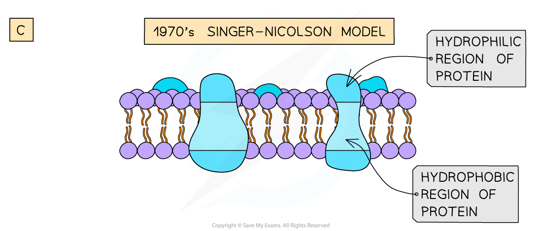
1970's Singer and Nicolson
- Singer and Nicolson proposed the fluid mosaic model which stated that membranes were fluid and that the globular proteins were both peripheral and integral (with some crossing both membranes) and dispersed throughout the membrane
- Evidence for this model:
- Analysis of freeze-etched electron micrographs showed proteins extending into the centre of membranes
- Biochemical analysis of the plasma membrane components
- The use of coloured fluorescent markers of antibodies. Antibodies were tagged with red and green fluorescent markers. These antibodies were bound to membrane proteins on different cells. Forty minutes after these cells were fused together the markers were seen to have mixed throughout the fused cells membrane showing that membrane proteins are free to move within the layer


Three models of membrane structure
Future models
- With further developments in technology more is still to be discovered about the plasma membrane and so the model we use to represent it continues to evolve
- e.g. the presence of the cellular cytoskeleton on the inside and the extracellular matrix on the outside makes the membrane less fluid than suggested by the fluid mosaic model
Exam Tip
You will need to learn the difference between the Davson-Danielli and Singer-Nicolson model of membrane structure and the reasons why they proposed their models.