DP Chemistry Questionbank

B.2 Proteins and enzymes
| Path: |
Description
[N/A]Directly related questions
-
16N.3.sl.TZ0.10b:
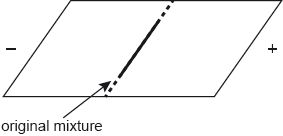
A mixture of amino acids is separated by gel electrophoresis at pH 6.0. The amino acids are then stained with ninhydrin.
(i) On the diagram below draw the relative positions of the following amino acids at the end of the process: Val, Asp, Lys and Thr.
(ii) Suggest why glycine and isoleucine separate slightly at pH 6.5.
-
16N.3.sl.TZ0.10d:
The fibrous protein keratin has a secondary structure with a helical arrangement.
(i) State the type of interaction responsible for holding the protein in this arrangement.
(ii) Identify the functional groups responsible for these interactions.
- 16N.3.sl.TZ0.10c: Determine the number of different tripeptides that can be made from twenty different amino acids.
-
17M.3.sl.TZ1.13b.i:
Identify the name of the amino acid that does not move under the influence of the applied voltage.
-
17M.3.sl.TZ1.13b.ii:
Deduce, giving a reason, which amino acid will develop closest to the negative electrode.
-
17M.3.sl.TZ1.13c:
The breakdown of a dipeptide in the presence of peptidase was investigated between 18 °C and 43 °C. The results are shown below.
Comment on the rate of reaction at temperature X in terms of the enzyme’s active site.
-
17M.3.sl.TZ2.7a:
Deduce the structural formula of the dipeptide Cys-Lys.
-
17M.3.sl.TZ2.7b:
Identify the type of bond between two cysteine residues in the tertiary structure of a protein.
-
17M.3.sl.TZ2.7c:
Deduce the structural formula of the predominant form of cysteine at pH 1.0.
-
17M.3.sl.TZ2.7d:
A mixture of the three amino acids, cysteine, glutamine and lysine, was placed in the centre of a square plate covered in polyacrylamide gel. The gel was saturated with a buffer solution of pH 6.0. Electrodes were connected to opposite sides of the gel and a potential difference was applied.
Sketch lines on the diagram to show the relative positions of the three amino acids after electrophoresis.

-
17M.3.hl.TZ2.8c.i:
An aqueous buffer solution contains both the zwitterion and the anionic forms of alanine. Draw the zwitterion of alanine.
-
20N.3.sl.TZ0.5b:
Proteins are polymers of amino acids.
Glycine is one of the amino acids in the primary structure of hemoglobin.
State the type of bonding responsible for the α-helix in the secondary structure.
-
20N.3.sl.TZ0.5a(ii):
Proteins are polymers of amino acids.
The mixture is composed of glycine, , and isoleucine, . Their structures can be found in section 33 of the data booklet.
Deduce, referring to relative affinities and , the identity of A1.
-
20N.3.sl.TZ0.5c:
Proteins are polymers of amino acids.
Describe how the tertiary structure differs from the quaternary structure in hemoglobin.
-
20N.3.sl.TZ0.5a(i):
Proteins are polymers of amino acids. A paper chromatogram of two amino acids, A1 and A2, is obtained using a non-polar solvent.
© International Baccalaureate Organization 2020.
Determine the value of A1.
-
20N.3.hl.TZ0.6a(ii):
Proteins are polymers of amino acids.
The mixture is composed of glycine, , and isoleucine, . Their structures can be found in section 33 of the data booklet.
Deduce, referring to relative affinities and , the identity of A1.
-
20N.3.hl.TZ0.6b:
Proteins are polymers of amino acids.
Glycine is one of the amino acids in the primary structure of hemoglobin.
State the type of bonding responsible for the α-helix in the secondary structure.
-
20N.3.hl.TZ0.6a(i):
Proteins are polymers of amino acids.
A paper chromatogram of two amino acids, A1 and A2, is obtained using a non-polar solvent.
© International Baccalaureate Organization 2020.
Determine the value of A1.
- 17N.3.sl.TZ0.11: Enzyme activity depends on many factors. Explain how pH change causes loss of activity of an enzyme.
-
18M.3.hl.TZ2.8c:
Draw the structures of the main form of glycine in buffer solutions of pH 1.0 and 6.0.
The pKa of glycine is 2.34.
-
18M.3.sl.TZ1.6a:
Draw the structural formula of a dipeptide containing the residues of valine, Val, and asparagine, Asn, using section 33 of the data booklet.
-
18M.3.sl.TZ1.6b:
Deduce the strongest intermolecular forces that would occur between the following amino acid residues in a protein chain.
-
18M.3.sl.TZ1.6c.ii:
Outline how the amino acids may be identified from a paper chromatogram.
-
18M.3.sl.TZ2.7a:
Draw the dipeptide represented by the formula Ala-Gly using section 33 of the data booklet.
-
18M.3.sl.TZ2.7c:
Outline why amino acids have high melting points.
- 18N.3.sl.TZ0.6a: Describe the interaction responsible for the secondary structure of a protein.
- 18N.3.hl.TZ0.8b: Explain the action of an enzyme and state one of its limitations.
- 18N.3.sl.TZ0.6b.i: Explain the action of an enzyme and state one of its limitations.
- 18N.3.hl.TZ0.8a: Describe the interaction responsible for the secondary structure of a protein.
-
19M.3.hl.TZ1.9a:
Draw a circle around the functional group formed between the amino acids and state its name.
Name:
-
19M.3.hl.TZ1.9b:
A mixture of phenylalanine and aspartic acid is separated by gel electrophoresis with a buffer of pH = 5.5.
Deduce their relative positions after electrophoresis, annotating them on the diagram. Use section 33 of the data booklet.
-
19M.3.hl.TZ2.9a(i):
Some proteins form an α-helix. State the name of another secondary protein structure.
-
19M.3.hl.TZ2.9a(ii):
Compare and contrast the bonding responsible for the two secondary structures.
One similarity:
One difference:
-
19M.3.hl.TZ2.9b:
Explain why an increase in temperature reduces the rate of an enzyme-catalyzed reaction.
-
19M.3.sl.TZ1.8a:
Draw a circle around the functional group formed between the amino acids and state its name.
Name:
-
19M.3.sl.TZ1.8b:
A mixture of phenylalanine and aspartic acid is separated by gel electrophoresis with a buffer of pH = 5.5.
Deduce their relative positions after electrophoresis, annotating them on the diagram. Use section 33 of the data booklet.
-
19M.3.sl.TZ2.6a(i) :
Some proteins form an α-helix. State the name of another secondary protein structure.
-
19M.3.sl.TZ2.6a(ii):
Compare and contrast the bonding responsible for the two secondary structures.
One similarity:
One difference:
-
19M.3.sl.TZ2.6b:
Explain why an increase in temperature reduces the rate of an enzyme-catalyzed reaction.
- 19N.3.hl.TZ0.10b(ii): Suggest why alanine and glycine separate slightly at pH 6.5.
- 19N.3.sl.TZ0.8a: The graph shows the relationship between the temperature and the rate of an enzyme-catalysed...
-
19N.3.sl.TZ0.7b:
The isoelectric point of amino acids is the intermediate pH at which an amino acid is electrically neutral.
Suggest why Asp and Phe have different isoelectric points.
-
19N.3.sl.TZ0.7a:
Draw the structure of the dipeptide Asp–Phe using section 33 of the data booklet.
-
19N.3.sl.TZ0.8b:
Explain why a change in pH affects the tertiary structure of an enzyme in solution.
- 19N.3.hl.TZ0.10b(i): Describe, using another method, how a mixture of four amino acids, alanine, arginine, glutamic...
-
19N.3.hl.TZ0.10a:
Draw the structure of the dipeptide Asp–Phe using section 33 of the data booklet.
