| Date | May 2018 | Marks available | 3 | Reference code | 18M.3.HL.TZ2.15 |
| Level | Higher level | Paper | Paper 3 | Time zone | Time zone 2 |
| Command term | Compare | Question number | 15 | Adapted from | N/A |
Question
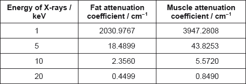
The attenuation values for fat and muscle at different X-ray energies are shown.

Outline the formation of a B scan in medical ultrasound imaging.
State what is meant by half-value thickness in X-ray imaging.
A monochromatic X-ray beam of energy 20 keV and intensity I0 penetrates 5.00 cm of fat and then 4.00 cm of muscle.

Calculate, in terms of I0, the final beam intensity that emerges from the muscle.
Compare the use of high and low energy X-rays for medical imaging.
Markscheme
many/array of transducers send ultrasound through body/object
B scan made from many A scans in different directions
the reflection from organ boundaries gives rise to position
the amplitude/size gives brightness to the B scan
2D/3D image formed «by computer»
[3 marks]
the thickness of tissue that reduces the intensity «of the X-rays» by a half
OR
\({x_{\frac{1}{2}}} = \frac{{\ln 2}}{\mu }\) where \({x_{\frac{1}{2}}}\) is the half value thickness and μ is attenuation coefficient
Symbols must be defined for mark to be awarded
[1 mark]
after fat layer, Ifat = I0e–0.4499 × 5.00
after muscle layer, I = Ifate–0.8490 × 4.00
I = 0.003533 I0 or 0.35%
[3 marks]
«high energies factors:»
less attenuation/more penetration
more damage to the body
«so» stronger signal leaves the body
OR
«so» used in «most» medical imaging techniques
«low energy factors:»
must be used with enhancement techniques
greater attenuation/less penetration
«so» more damage to the body «on surface layers»
OR
«so» unwanted in «most» medical imaging techniques
[3 marks]

